| | Hypothalamus | Anterior pituitary | Ovary | Oviduct | Uterus | Vagina | Vulva and clitoris | Ovarian Reproductive Functions and Estrous Cycle
The purpose of the information presented in this section is to familiarize the reader with the terminology, anatomical location and functional anatomy of the important components of the cow's reproductive system.
Hypothalamus
The hypothalamus is an organ located within the brain and is considered to be the coordinating center for most of the reproductive activities and changes in mammalians. Numerous neural cell bodies located within the hypothalamus are responsible for producing in a pulsatile manner (pulse generator) the Gonadotropin Releasing Hormone (GnRH), which is the hormone responsible for controlling the whole reproductive system (see Fig.2).
After a short activity in the first days of life, the GnRH pulse generator enters in a state of dormancy until a few days prior to puberty, when the heifer is entering sexual maturity. In that point, the GnRH pulse generator system is activated and GnRH is released from the hypothalamus into the capillary system (hypophyseal-portal system) which connects the hypothalamus with the anterior pituitary. Factors from both within the animal and from the external environment may affect the amount and pattern of GnRH pulse generator and release by the hypothalamus, which in the cow could include temperature, photoperiod, stress (social, behavioral, disease, nutrition), the use of certain medications, such as opioids, and stage of the reproductive cycle (estrus, pregnancy, parturition, suckling).
Anterior Pituitary
The pituitary is a gland located at the base of the brain and is connected to the hypothalamus via the hypophyseal-portal system. The anterior lobe of the pituitary gland has a critical role in reproduction, which involves the production and release of the two gonadotropic hormones: luteinizing hormone (LH) and follicle-stimulating hormone (FSH) (see Fig.2). Since the GnRH is released in a pulsatile manner, it triggers the release of both, LH and FSH in pulses, which in the case of LH may occur at a frequency of one pulse per hour once puberty has been reached. After release from the anterior pituitary, LH and FSH travel through the animal's general circulatory system to reach the ovaries. LH pulses result on production of progesterone, an ovarian hormone, which prepares the initial stages for follicular development. As the cycle progresses, another ovarian hormone, estradiol, which is produced as a result of FSH stimulation, progressively increases its concentration in the bloodstream until it causes a major LH surge that results in ovulation. Then, the cycle is repeated with the post ovulatory ovary beginning its production of progesterone, under LH influence. Production and release of LH and FSH can be directly influenced by negative feedback from ovarian hormones, or indirectly by the same factors which can alter hypothalamic function.
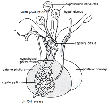
Fig.2. The hypothalamus is located in the brain and is connected to the pituitary gland through the hypophyseal portal system (vessels). GnRH is produced in neuronal cells of the hypothalamus and transported to the pituitary through the hypophyseal portal vessels to stimulate the production of LH and FSH in the anterior pituitary gland. These pituitary hormones are released to the general circulation in a pulsatile manner in different concentrations, and have an effect mostly on the ovary to stimulate the production of ovarian hormones, which are depending on the stage of the estrus cycle.
Ovary
The normal, mature cow possesses two almond shaped ovaries, 1 to 4 cm long and 1 to 3 cm wide. The ovaries are suspended in the abdominal cavity by a ligament (the mesovarium) and are partially surrounded by the infundibulum (Fig 1).
Oviduct
The oviduct is a coiled tube which transports the newly ovulated ovum to the point of fertilization and down to the uterus for implantation. In the cow, the oviduct is about 10 inches (25 cm) long when fully stretched out (Fig.1). From a functional point of view, the oviduct is made up of three parts:
1. The closest area to the ovary is the infundibutum with its finger-like projections called fimbria. The only attachment between the oviduct and ovary occurs at one point of the fimbria and the upper pole of the ovary.
2. the ampulla connects at the ovarian end with the infundibulum and at its distal end with the isthmus. The ampulla-isthmus junction is the point where fertilization of the ovum takes place.
3. the isthmus is connected directly with the uterine horn.
The oviducts are made up of ciliated cells, secretory cells and smooth muscle cells. The ciliated cells, present in greater abundance in the fimbria and infundibulum and decreasing in concentration towards the uterus, move the ovum from the ovary towards the uterus. The activity of the ciliated cells, coupled with contractions of the smooth muscle cells, keeps an ovulated ovum in constant rotation which is essential for bringing egg and sperm together and preventing oviductal implantation. The secretory cells are responsible for producing mucous like substances which coat and protect the ovum. The activity of the cells in the oviduct is controlled primarily by estrogen produced by the ovary and oxytocin released by the posterior pituitary.
Uterus
The uterus of the cow is suspended in the body cavity by the broad ligaments which attach to the pelvic and abdominal walls. The uterus consists of two long horns, a relatively short body and a neck or cervix (Fig.1). The uterus performs a number of functions:
- Sperm is transported from the vagina to the oviduct during heat and the fetus is expelled at term by uterine muscle contractions;
- Sperm capacitation, which is essential for fertilization, occurs when sperm pass through uterine secretions;
- Fertilized ovum (embryos) are provided nourishment before implantation by the uterine secretions; and
- Prostaglandins are produced by the uterus which can destroy the function of the corpus luteum of the ovary.
The inner surface of the uterus in the cow also contains 70 to 120 caruncles through which the fetal placenta attaches and the fetus receives nourishment during pregnancy. Muscular and secretory activity of the uterus is controlled by ovary hormones and oxytocin from the posterior pituitary.
The cervix is the neck of the uterus which connects the body of the uterus with the vagina. The cervix is characterized by a thick wall and constricted lumen with interlocking, transverse rings (usually four rings in the ciliated cells beat towards the vagina to remove debris and secretory cells produce mucous-like substances to either aid in sperm transport or act as a block further invasion of organisms coming from the vagina, depending on the stage of the reproductive cycle. The primary function of the cervix is to prevent intruders from entering the uterus. The cervix is tightly closed except during estrus at which time R relaxes slightly allowing rm to enter the uterus. During pregnancy, an increased amount of very viscous cervical fluid blocks the cervical canal and acts to prevent uterine infection. The cervix opens again just before calving and the mucous lug liquefies, allowing delivery of the fetus and fetal membranes. Like the rest of the uterus, the activity of the cervix is controlled by ovarian hormones.
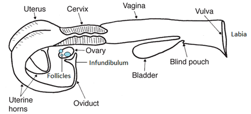
Fig. 1. Diagrammatic sketch of the reproductive tract of the cow. The follicles are located in the ovary and are round structures in different stages of development filled with follicular fluid (blue) containing different concentrations of estradiol and other hormones.
Vagina
The vagina is an elastic tube connecting the external opening of the vagina with the cervix (Fig.1). The vagina functions as a receptacle for the male penis during natural service and as a passageway for the fetus and fetal membranes during parturition. Mucous-secreting cells of the vagina provide lubrication for both the penis and the fetus. Although not as well developed as in the uterus, a muscle layer is present in the vagina and may aid in sperm transport and fetal expulsion. The urinary urethra also opens into the posterior portion of the vagina and both urine and reproductive secretions exit the vagina through this common pathway.
Vulva and clitoris
The vulva is the external opening of the vagina through which urine, uterine and vaginal secretions are excreted and the fetus and fetal membranes are expelled at term (Fig.1). On the floor of the vagina just inward from the vulva, the clitoris is situated. The clitoris, although considerably smaller, is similar to the male penis and is well supplied with sensory nerve endings. The clitoris is stimulated naturally by the bull during mating and if stimulated manually during artificial insemination, it can result in increased conception rates in mature cows. Clitoral stimulation is believed to cause an increase in uterine contractions such that sperm transport is enhanced.
Ovarian Reproductive Functions and Estrous Cycle
The estrus cycle is an event that occurs periodically in sexually mature cows and it is controlled by the hypothalamo-pituitary-ovarian axis and creates the opportunity for mating and reproduction of the specie.
The ovary has two primary functions:
a) Production and release of the female reproductive cell, the egg (ovum, or oocyte); and
b) Production and release of ovarian hormones (estrogen, progesterone, inhibin, oxytocin and relaxin).
The oocyte is contained within a small, blister-like structure called the follicle and is surrounded by follicular fluid and the cells responsible for estrogen production (the granulosa cells) forming the cumulus-oocyte complex. The cow is born with the maximum number of immature ova (termed oocytes) that she will ever have (between 60,000 and 80,000).
Numerous immature, oocyte-containing primordial follicles enter the growing phase throughout the cow's reproductive lifetime, but the cow generally releases only one ovum into the infundibulum at ovulation. The follicles which fail to ovulate undergo death or atresia and degeneration. Factors determining which of the ovarian oocytes are destined to begin their growth or to complete their growth during a reproductive cycle remain unknown. There are two stages in the growth of the oocyte.
During the first stage, oocyte growth is rapid and closely associated with development of the ovarian follicle. The oocyte attains its mature size by the end of this first growing phase. This first growing phase also does not appear to be controlled by the pituitary hormones, FSH and LH.
During the second stage of the growth cycle, the oocyte does not grow in size, but the ovarian follicle increases in diameter very rapidly and begins secreting estrogen in significant quantities. This second stage of growth is under the control of the pituitary hormones, LH and FSH.
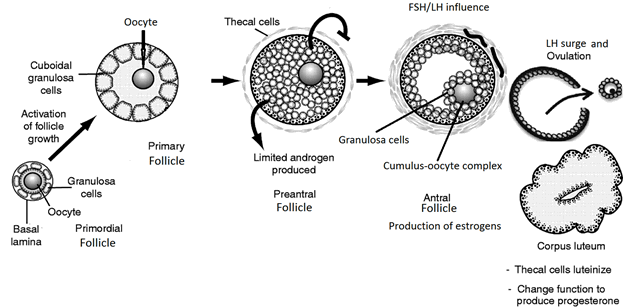
Fig.3. Different stages of follicular growth in mature cows. Pre-antral follicles develop under the influence of both, FSH and LH pulses. Antral (pre-ovulatory and ovulatory follicles) have increased production of estrogens from the granulosa cells, causing the behavioral heat or estrus, which lasts from 12-18 hours. These high estrogen concentrations trigger a surge in LH, which results in ovulation that typically occurs 24-30 hours after the onset of heat. The ovulatory follicle becomes luteinized under the influence of LH, with increased production of progesterone. If pregnancy occurs, progesterone helps maintain pregnancy during most of the gestation.
After ovulation and release of the egg from the follicle, the cells remaining in the follicle shell begin rapid division and change or differentiation from estrogen-producing cells to progesterone-producing cells (Fig.3). The progesterone-producing cells are termed luteal cells and they make up the yellow body or corpus luteum (CL) of the ovary. The CL produces progesterone in response to LH stimulation. If the cow is not pregnant, the CL dies or regresses in response to a hormone (prostaglandin F2�) secreted by the uterus and progesterone production declines rapidly by about day 16 to 18 of the estrous cycle. The estrous cycle itself last about 3 weeks, but may range from 17 to 25 days.
If the cow is pregnant, the CL remains active until parturition. In addition to secreting progesterone, the CL of pregnancy is also responsible for the production and secretion of relaxin, which causes cervical and pelvic relaxation at term. Some cells within the CL also produce the hormone oxytocin, which may be involved in luteal regression and termination of the luteal phase in a regular estrus cycle, and together with prostaglandin F2� and fetal cortisol, are involved in the termination of pregnancy and on the parturition process.
Adapted from Beef Herd Management Reference Binder and Study Guide 403-1
|
|