| | General life cycle of coccidia | Special features of the life cycle | How do birds become infected? | How do coccidia harm chickens? | How to know when chickens are infected? | How to prevent chickens from getting coccidiosis | How to be sure chickens have coccidia | How to treat infected birds
Coccidiosis causes considerable economic loss in the poultry industry. Chickens are susceptible to at least 11 species of coccidia.
The most common species are Eimeria tenella, which causes the cecal or bloody type of coccidiosis, E. necatrix, which causes bloody intestinal coccidiosis, and E. acervulina and E. maxima, which cause chronic intestinal coccidiosis.
General Life Cycle of Coccidia
Stages of coccidia in chickens appear both within the host as well as outside. The developmental stages in the chicken give rise to a microscopic egg (called an oocyst) that is passed out in the droppings.
Under proper conditions of temperature and moisture, the oocyst develops within one to two days to form a sporulated oocyst, which is capable of infecting other chickens. At this stage, the oocyst contains eight bodies (called sporozoites), each of which is capable of entering a cell in the chicken's intestine after the oocyst is eaten.
When sporozoites enter the cells, they divide many times producing either a few or many offspring (merozoites). The numbers produced depend on the species of coccidia involved. Each merozoite, in turn, may enter another intestinal cell. This cycle may be repeated several times. Because of this cyclic multiplication, large numbers of intestinal cells are destroyed.
Eventually, the cycle stops and sex cells (male and female) are produced. The male fertilizes the female to produce an oocyst, which ruptures from the intestinal cell and passes in the droppings. Thousands of oocysts may be passed in the droppings of an infected chicken; therefore, poultry raised in crowded or unsanitary conditions are at great risk of becoming infected.
Special Features of the Life Cycle
Eimeria tenelia
This parasite develops in the cells of the ceca, which are the two blind sacs near the end of the intestine. It is one of the most pathogenic (disease producing) coccidia to infect chickens. This acute infection occurs most commonly in young chicks. Infections may be characterized by the presence of blood in the droppings and by high morbidity and mortality.
E. necatrix
E. necatrix develops in the small intestine (early stages) and later in the cecum (sexual stages). Like E. tenella, it develops within deeper tissues of the small intestine and is a major pathogen of poultry.
E. acervulina and E. maxima
Both species develop in epithelial cells, primarily in the upper part of the small intestine. They cause subclinical coccidiosis associated with marked weight loss.
How do Birds Become Infected?
Normally, most birds pass small numbers of oocysts in their droppings without apparent ill effects. Coccidiosis becomes important as a disease when animals live, or are reared, under conditions that permit the build-up of infective oocysts in the environment. The intensive rearing of domestic chickens may provide these conditions.
Young chickens pick up the infection from contaminated premises (soil, houses, utensils, etc.). These premises may have been contaminated previously by other young infected birds or by adult birds that have recovered from the condition. Wet areas around water fountains are a source of infection.
Oocysts remain viable in litter for many months. In this way, they can contaminate a farm from year to year. Oocysts are killed by freezing, extreme dryness and high temperatures.
How do Coccidia Harm Chickens?
Several factors influence the severity of infection. Some of these include the following:
- The number of oocysts eaten. Generally, an increase in the number of oocysts eaten is accompanied by an increase in the severity of the disease.
- Strain of coccidia. Different strains of a species may vary in pathogenicity.
- Environmental factors affecting the survival of the oocysts.
- Site of development within the host. Coccidia that develop superficially are less pathogenic than those that develop deeper.
- Age of the bird. Young birds are generally more susceptible than older ones.
- Nutritional status of the host. Poorly fed birds are more susceptible.
Coccidiosis in chickens is generally classified as either intestinal or cecal. Most serious cases of intestinal coccidiosis are caused by E. necatrix. Cecal coccidiosis is due to E. tenella.
Coccidiosis occurs most frequently in young birds. Old birds are generally immune as a result of prior infection. Severe damage to the ceca and small intestine accompany the development of the coccidia. Broilers and layers are more commonly infected, but broiler breeders and turkey and pheasant poults are also affected.
Coccidiosis generally occurs more frequently during warmer (May to September) than colder months (October to April) of the year. E. acervulina and E. maxima develop in epithelial cells within the small intestine and generally cause chronic intestinal coccidiosis.
How to Know When Chickens are Infected?
The most easily recognized clinical sign of severe cecal coccidiosis is the presence of bloody droppings. Dehydration may accompany cecal coccidiosis.
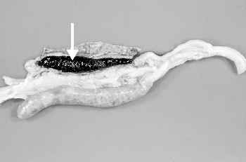
Coccidiosis caused by E. tenella first becomes noticeable at about three days after infection. Chickens droop, stop feeding, huddle together, and by the fourth day, blood begins to appear in the droppings. The greatest amount of blood appears by day five or six, and by the eighth or ninth day, the bird is either dead or on the way to recovery. Mortality is highest between the fourth and sixth days. Death may occur unexpectedly, owing to excessive blood loss. Birds that recover may develop a chronic illness as a result of a persistent cecal core. However, the core usually detaches itself by eight to ten days and is shed in the droppings.

Figure 1. Coccidiosis caused by E.tenella developing in the ceca. The arrow indicates the cecal core
E. necatrix causes a more chronic disease than E. tenella and does not produce as many oocysts. Therefore, a longer time is usually required for high levels of environmental contamination. Birds heavily infected with E. necatrix may die before any marked change is noticed in weight or before blood is found in the feces.
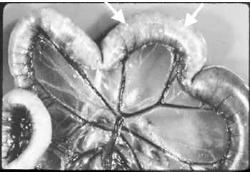
E. acervulina is less pathogenic than E. tenella or E. necatrix. E. acervulina is responsible for subacute or chronic intestinal coccidiosis in broilers, older birds and chickens at the point of lay. The clinical signs consist of weight loss and a watery, whitish diarrhea. At postmortem, greyish-white, pinpoint foci or transversely elongated areas are visible from the outer (or serous) surface of the upper intestine. The foci consist of dense areas of oocysts and gamete (male and female sex cells) production.

Figure 2. Greyish-white foci in the intestine caused by E. necatrix
E. maxima produces few marked changes in the small intestine until the fifth day after infection. After that time in severe infections, numerous small hemorrhages occur along with a marked production of thick mucus. The intestine loses tone and becomes flaccid and dilated. The inner surface is inflamed, and the intestinal content consists of a pinkish mucoid secretion.
How to Prevent Chickens from Getting Coccidiosis
A few good management practices will help control coccidiosis. Contact your veterinarian for full details.
- Anticoccidial drugs mixed in the feed are used to limit high levels of infection.
- Keep chicks, feed and water away from droppings.
- Roost birds over wire netting if brooding arrangements make this possible.
- Place water vessels on wire frames to eliminate a concentration of wet droppings, in which the chicks can walk to pick up or spread the disease.
- Keep litter dry and stirred frequently. Remove wet spots and replace with dry litter.
- Avoid overcrowding.
- If coccidiosis does break out, start treatment immediately.
How to be sure chickens have Coccidia
Diagnosis is best accomplished by postmortem examination of a representative number of birds from the flock. The location of the major lesions gives a good indication of the species of coccidia concerned.
For example, hemorrhagic lesions in the central part of the small intestine suggest E. necatrix, those in the cecum, E. tenella. Microscopic examination of the affected areas along with measuring oocysts will confirm the identification.
How to Treat Infected birds
Several anticoccidials are currently available. Depending on the product used, the withdrawal periods and contraindications should be strictly followed.
The emergence of drug-resistant strains of coccidia may present a major problem. Methods used to avoid the development of drug resistance include switching classes of drugs and the "shuttle program," which is a planned switch of drug in the middle of the bird's growing period.
Source: Agdex 663-35. Revised April 2001. |
|