| | Background
Whether you are a table egg producer, a turkey or broiler breeder egg producer, or someone who grows out turkeys or broilers for processing, in one way or another, your livelihood depends on eggs. Poultry people have a greater appreciation for eggs than most folks who simply enjoy eggs with a side of bacon, but have you really thought about all the different parts of an egg and what their purpose is?
What is an egg?
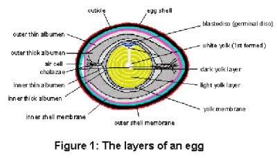
All bird eggs provide a self-contained package in which growth of the embryo (developing young bird) occurs. Unlike humans, embryo growth in birds occurs outside of the body of the female, and therefore, all of the solid nutrients required for the embryo to grow and develop must be present in the egg at the time it is laid. It is the nutrients in the diet of the hen which eventually form the different parts of the egg. Figure 1 shows a picture of an egg as if the egg were cut lengthwise through the middle. The various parts of the egg will be described in the approximate order in which they are formed in the reproductive tract of the hen.
Blastodisc (Germinal Disc)
The blastodisc is a single cell that originates from the hen and is positioned on the surface of the yellow yolk. Penetration of sperm from the rooster into the blastodisc completes fertilization. Once fertilized, the blastodisc divides and grows and is now called a blastoderm (early embryo). It is only this small group of cells which divide and develop; the large mass of yolk upon which the embryo sits does not divide and only serves to provide food to the embryo.
Yolk
The yolk is the primary food source for the growing embryo. The yolk follicle is formed on the ovary of the hen. As a percentage of the yolk weight, yolk is made up of mostly water, then fat (lipids), then proteins. The first yolk that is formed is white in color. Because it is the first yolk formed, it takes up a position in the centre of the yolk follicle. This small portion of white yolk in the centre of the yellow yolk provides the first nutrients for the early embryo to grow and develop. The yellow yolk is placed in alternating dark and light layers, one on top of another.
Yolk membrane
The yolk membrane is the structure that surrounds and contains the yolk and the early embryo positioned on top of the yolk. It is made up of several layers formed while the yolk is on the ovary and after the yolk is ovulated (released from the ovary into the reproductive tract). The two main layers of the yolk membrane consist mostly of a protein that produces a mesh-like structure.
Albumen (Egg White)
In the egg there are two types of albumen: thin and thick. Both types of albumen are made up mostly of proteins and water. The inner thick albumen is the portion in contact with the yolk membrane surrounding the yolk. Its function is to cushion the yolk and embryo and to inhibit the entry of bacteria into the yolk and embryo area. Some of the other proteins found in both types of albumen act to prevent entry of bacteria or to kill bacteria. Other proteins in the albumen contribute protein building blocks (amino acids) for embryo growth.
Chalazae (singular - chalaza)
The chalazae are ropelike structures that are made up of the same types of proteins found in the thick albumen. The chalazae function to connect the yolk and inner thick albumen to the outer thick albumen at the long axis of the egg. This stabilizes the yolk in the centre of the egg and prevents the yolk and embryo from hitting the inside of the shell when the egg is moved. The chalaza at the small end of the egg consist of two twisted strands of thick albumen-like material, while the chalaza at the large end of the egg consists of only one strand.
Shell membranes
There are two shell membranes present in an egg; the inner shell membrane, which is in contact with the albumen, and the outer shell membrane, which is in contact with the shell. The shell membranes are closely adhered to each other except at the large end of the egg where they separate to form the air cell. Both shell membranes are made up of protein fibers that form a mesh-like structure to hold the albumen in place and provide a barrier to entry of bacteria.
Air cell
The air cell is a pocket of air that forms at the large end of the egg. As the egg loses moisture during storage or throughout incubation, the air cell grows in size. During the hatching process, the chick embryo penetrates through the inner shell membrane with its beak and into the air cell to obtain its first breath.
Egg shell
The hard shell is made up of several layers and is mostly composed of the compound calcium carbonate. The final layer of shell contains the egg shell pigments. In wild birds, the shell pigments provide camouflage to hide eggs and protect them from predators. The obvious function of the shell is to contain the egg components and to provide a protective barrier against bacteria and physical shock for the developing embryo. The shell also provides calcium and other minerals to the growing embryo. Although the shell appears to be a solid structure, there are small pores (holes) in the shell which allow the transfer of gases into and out of the egg. Oxygen is transferred through the shell into the egg, and is used by the growing embryo to breathe. When the embryo uses the oxygen, carbon dioxide and water vapor are produced and then transferred out of the egg through the shell.
Egg shell cuticle
When the egg is first laid the shell is wet. As the shell dries, a hard thin covering over the shell is formed. This layer, mostly consisting of protein, is called the cuticle. The function of the cuticle is to prevent water loss from the egg and entry of bacteria into the egg. The cuticle is the first line of defense against bacteria. When hatching eggs are washed, scrubbed, or cleaned using sandpaper, this protective coating is removed and increases the chance of
bacteria entry into the egg.
G. M. Fasenko, University of Alberta
Poultry Research Centre News - Vol. 8 No. 1, March 1998 |
|